Embark on a journey into the intricate realm of neuroanatomy with Gross Anatomy of the Brain and Cranial Nerves Exercise 17. This comprehensive exploration unveils the remarkable structures and functions of the central nervous system, providing a profound understanding of its significance in human health and well-being.
Through detailed descriptions, engaging illustrations, and practical dissection guidance, this exercise delves into the intricacies of the brain, from its protective meninges to its specialized regions, and the enigmatic cranial nerves that connect it to the rest of the body.
Prepare to unravel the complexities of the brain and cranial nerves, gaining invaluable insights into their clinical applications and the neurological disorders they impact.
Gross Anatomy of the Brain

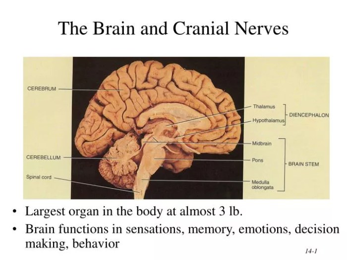
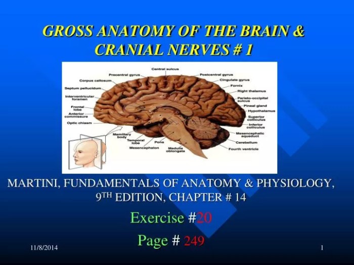
The brain is the central organ of the nervous system and is responsible for controlling most bodily functions. It is divided into three main parts: the cerebrum, cerebellum, and brainstem.
Cerebrum
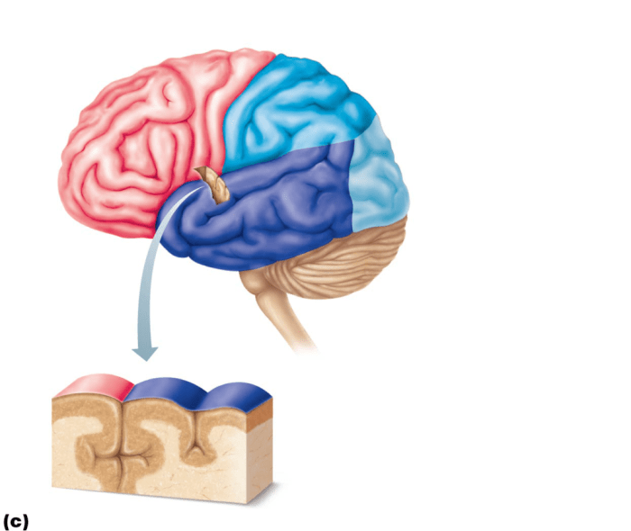
The cerebrum is the largest part of the brain and is responsible for higher-level functions such as cognition, language, and memory. It is divided into two hemispheres, the left and right hemispheres, which are connected by the corpus callosum.
Cerebellum, Gross anatomy of the brain and cranial nerves exercise 17
The cerebellum is located at the back of the brain and is responsible for coordination and balance. It receives sensory input from the body and sends signals to the muscles to help coordinate movement.
Brainstem
The brainstem is located at the base of the brain and is responsible for vital functions such as breathing, heart rate, and blood pressure. It is divided into three parts: the midbrain, pons, and medulla oblongata.
Meninges and Cerebrospinal Fluid
The brain is surrounded by three layers of meninges, which are protective membranes. The meninges are filled with cerebrospinal fluid, which cushions the brain and provides nutrients.
Cranial Nerves

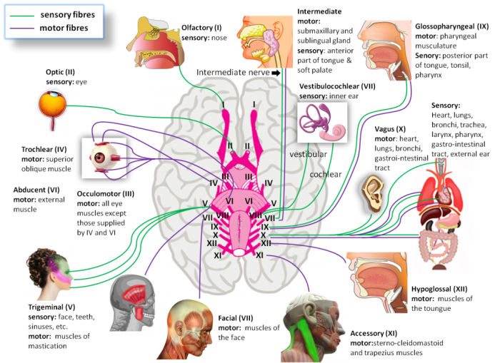
The cranial nerves are 12 pairs of nerves that connect the brain to the head and neck. They are responsible for a variety of functions, including vision, hearing, smell, taste, and movement.
List of Cranial Nerves
- Olfactory nerve (I)
- Optic nerve (II)
- Oculomotor nerve (III)
- Trochlear nerve (IV)
- Trigeminal nerve (V)
- Abducens nerve (VI)
- Facial nerve (VII)
- Vestibulocochlear nerve (VIII)
- Glossopharyngeal nerve (IX)
- Vagus nerve (X)
- Accessory nerve (XI)
- Hypoglossal nerve (XII)
Functions of Cranial Nerves
The cranial nerves are responsible for a variety of functions, including:
- Vision
- Hearing
- Smell
- Taste
- Movement of the eyes, face, and tongue
- Salivation
- Swallowing
- Heart rate
- Blood pressure
Exercise 17: Dissection of the Brain and Cranial Nerves

The dissection of the brain and cranial nerves is a complex procedure that requires careful dissection techniques. The following steps provide a general overview of the dissection process:
- Remove the scalp and skullcap to expose the brain.
- Identify the major structures of the brain, including the cerebrum, cerebellum, and brainstem.
- Dissect the meninges to expose the surface of the brain.
- Identify the cranial nerves and trace their course through the brain.
- Remove the brain from the skull and place it in a dissection tray.
- Dissect the brain into smaller sections to examine its internal structures.
The dissection of the brain and cranial nerves is a valuable learning experience that provides students with a detailed understanding of the anatomy of these structures.
Clinical Applications: Gross Anatomy Of The Brain And Cranial Nerves Exercise 17

The gross anatomy of the brain and cranial nerves is essential for understanding a variety of neurological disorders. For example, damage to the cerebrum can lead to problems with cognition, language, or memory. Damage to the cerebellum can lead to problems with coordination and balance.
Damage to the brainstem can lead to problems with vital functions such as breathing, heart rate, and blood pressure.
Knowledge of the gross anatomy of the brain and cranial nerves is also essential for performing a variety of medical procedures. For example, neurosurgeons use this knowledge to operate on the brain and cranial nerves. Neurologists use this knowledge to diagnose and treat neurological disorders.
Essential Questionnaire
What is the significance of the meninges in brain protection?
The meninges, consisting of the dura mater, arachnoid mater, and pia mater, provide crucial protection to the delicate brain tissue. They act as a physical barrier against mechanical trauma, infection, and hemorrhage, ensuring the optimal functioning of the central nervous system.
How do cranial nerves differ from spinal nerves?
Cranial nerves, unlike spinal nerves, originate directly from the brainstem and are responsible for innervating structures within the head and neck. They control a wide range of functions, including sensory perception, motor control, and autonomic regulation.
What are some common neurological disorders that affect the brain and cranial nerves?
Numerous neurological disorders can impact the brain and cranial nerves, including stroke, Parkinson’s disease, Alzheimer’s disease, and multiple sclerosis. These conditions disrupt the normal functioning of the central nervous system, leading to a variety of symptoms and impairments.